Fig. 5.
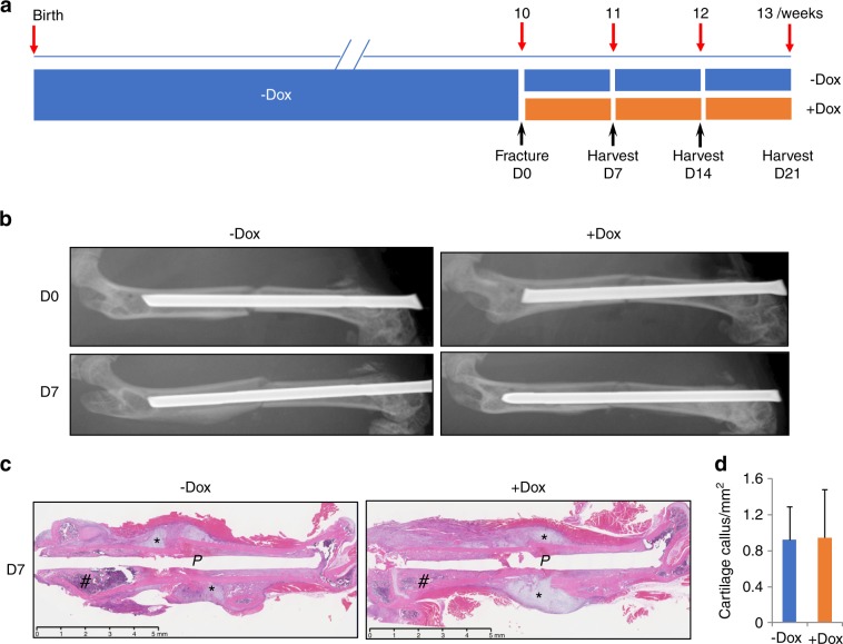
Wnt7b overexpression does not affect the cartilage callus of fractures. a A schematic of experimental design. b Representative X-ray images of femurs immediately following fracture (D0) and at 7 days after procedure (D7). c Representative H&E images of medial sections through the fractured femur at D7. P stabilizing pin, asterisk indicates cartilage callus. Note excessive bone accrual in distal metaphyseal region (#) of the +Dox mouse. d Measurements of cartilage callus areas on sections. N = 3 mice. Three sections per mouse were measured with the NanoZoomer NDP software.

