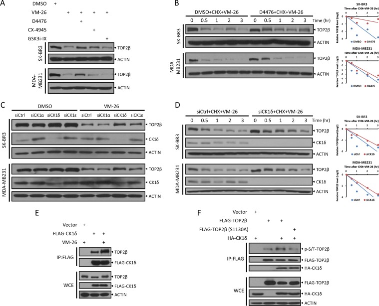
Fig. 5. CK1 binds with and phosphorylates TOP2β at Ser1130 to promote its degradation by VM-26.
a Inhibition of CK1, but not CK2 or GSK3, blocks the degradation of TOP2β induced by VM-26. Cells pretreated with D4476, CX-4945 or GSK3i-IX for 1 h were treated with VM-26 for an additional 2 h. Cells were then harvested for IB analysis with the indicated Abs. b Inhibition of CK1 extends the protein half-life of TOP2β. Cells left untreated or pretreated with D4476 (50 μM) for 4 h were then exposed to CHX and VM-26. Cells were harvested at the indicated time points for IB analysis with the indicated Abs (left). Densitometry quantification was performed with ImageJ, and the decay curves are shown (right). c and d Silencing of CK1 inhibits TOP2β degradation by extending its protein half-life. Cells transfected with the indicated siRNA oligos were treated with VM-26 for 1 h (c) or treated with VM-26 and CHX for the indicated time periods (d), and then, IB was undertaken with the indicated Abs. Densitometry quantification was performed with ImageJ, and the decay curves are shown (d, right). e, f CK1 binds to and phosphorylates TOP2β at Ser1130. HEK293 cells transfected with the indicated plasmids were treated with VM-26 for 5 h as indicated (e) or in combination with MG132 (f), and then, IP was conducted with anti-FLAG beads (top), and direct IB was undertaken with the indicated Abs (bottom).

