
Fig. 4. Recombinant αSyn fragments are readily taken-up by naïve LUHMES neurons.
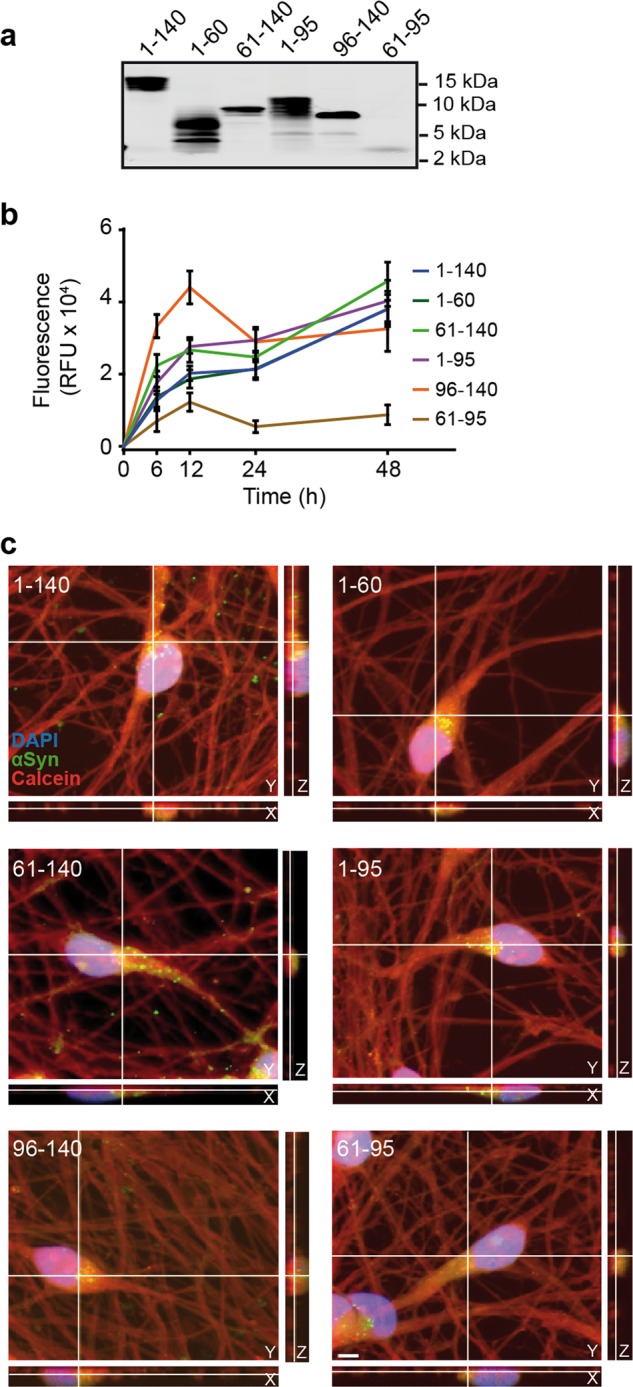
a Fluorescent gel imaging of recombinant ATTO-488-labeled FL-αSyn and fragments (depicted in Fig. 3a). All labeled αSyn species have a fluorescent signal at the expected molecular size. b Uptake kinetics of recombinant FL-αSyn and fragments. Cells were treated with labeled αSyn at DIV4. At the indicated times, intracellular fluorescence was measured. Extracellular fluorescence was quenched using trypan blue. RFU: relative fluorescence unit. c Uptake of ATTO-488-labeled αSyn fragments 48 h after treatment. Confocal images show orthogonal projections of Z-stacks with a clear intracellular localization of recombinant FL-αSyn and fragments. A calcein red-orange filling (red), DAPI staining (blue) and labeled recombinant αSyn (green) are shown. Scale bar: 5 µm.