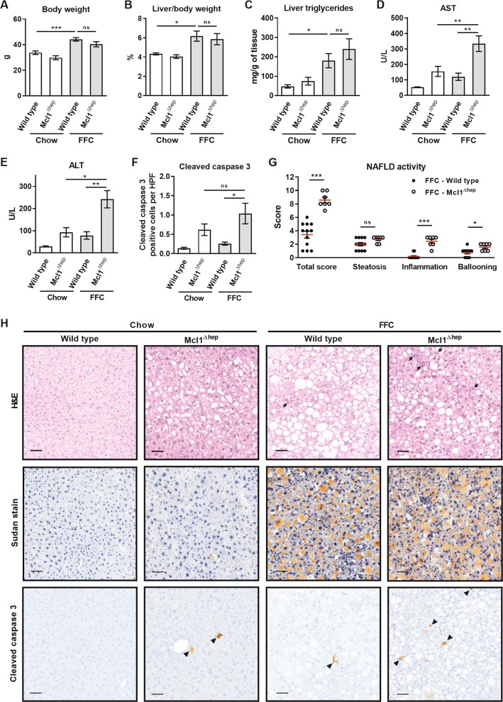
Fig. 2. Mcl1 deficiency in hepatocytes exacerbates liver injury induced by the FFC diet.
Mcl1∆hep mice and control littermates (WT) were placed on standard chow or FFC diet for 4 months. Blood and livers were harvested at the end of the study. a Body weight at the end of the feeding study. b Liver weight as percentage of total body weight. c Liver triglycerides were measured in tissue homogenates using biochemical assay. d, e Liver injury was assessed by serum AST and ALT activity. f Hepatic apoptosis was assessed by immunohistochemistry and counting cleaved caspase 3-positive cells per 20× fields. n = 5 mice/group. g NAFLD activity score. FFC-WT n = 12 mice; FFC-Mcl1∆hep n = 7 mice; h Representative images of H&E staining (arrows point to inflammatory foci), Sudan red staining for lipids, and immunohistochemistry for cleaved caspase 3 (arrowheads) in liver tissue samples (scale bar 50 μm). Bars represent mean ± SEM. a–e Chow-WT n = 9 mice; Chow-Mcl1∆hep n = 5 mice; FFC-WT n = 12 mice; FFC-Mcl1∆hep n = 7 mice; ***p < 0.001, **p < 0.01, *p < 0.05 or not significant (ns).

