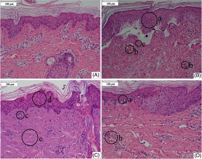
Fig. 5.

Histological sections of skin tissue. (A) Normal skin tissue. (B) Damaged skin tissue at 48 h post exposure. Laser conditions: beam diameter 1.96 cm, exposure duration 3.0 s, and laser power 16.0 W. The radiant exposure was about 1.11 times of the damage threshold. (C) Skin tissue at the central region of a damage lesion at 48 h post exposure. Laser conditions: beam diameter 1.96 cm, exposure duration 3.0 s, and laser power 24.0 W. The radiant exposure was about 1.67 times of the damage threshold. (D) Skin tissue at the edge region of the same lesion as (C). Laser condition was the same as (C). The specimens seen in (C) and (D) were from the same tissue section but different locations. The circles from (a) to (e) represented following damage characteristics: (a) gathered nuclear chromatin in the epidermis, (b) blood cells deposited abundantly in the capillary vessels, (c) the cell nuclei in the upper dermis layer shrank with hyperchromatism, (d) obvious stretching of the nuclear chromatin in the epidermis, and (e) the structure of the collagen fibers changed obviously with homogenization characteristic.
