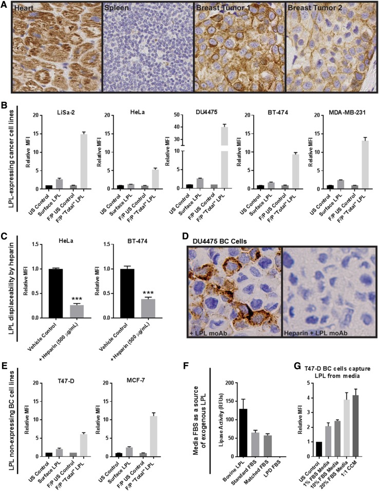
Fig. 1.
LPL is in and on the surface of breast tumor cells and cancer cell lines and is displaceable by heparin. A: Control (heart and spleen) and breast tumor slides were stained for LPL. Breast tumor cells display cell surface and cytoplasmic LPL staining. B: The presence of LPL protein in and on the surface of cancer cells was assessed via flow cytometry. For all cell lines, the total LPL present in fixed/permeabilized (F/P) cells exceeded that on the cell surface. Note different scales; mean ± SEM of >3 experiments. C: Representative data from heparin displacement studies using LPL-expressing HeLa cervical cancer and BT-474 BC cells. Heparin displaces LPL from the cell surface, as detected by flow cytometry (***P < 0.001; two-tailed unpaired t-test with Welch’s correction). D: Immunocytochemical analysis of LPL on the surface of DU4475 nonadherent TNBC cells. Cells were incubated with LPL antibody ± heparin (40 µg/ml), embedded in agarose, formalin-fixed, embedded in paraffin, and sectioned. Cell-surface LPL antibody was detected with peroxidase-conjugated anti-IgG (brown pigment) with hematoxylin counterstain (blue). E: T47-D and MCF-7 BC cells had no detectable LPL expression by qRT-PCR. However, LPL was found on the cell surface and, to a greater extent, inside these cells by flow cytometry. F: Standard, LPD, and matched-control FBS were assessed for lipase activity. Activity was detected in bovine LPL and in standard and matched-control FBS but not LPD FBS. G: T47-D BC cells were incubated in different media overnight to determine whether BC cells can capture LPL from media. A FBS dose-dependent increase of cell-surface LPL was observed. LPL was also captured by cells incubated with culture media conditioned by LPL-secreting LiSa-2 liposarcoma cells.

