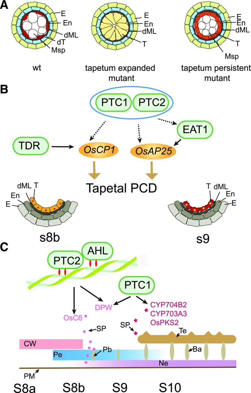
Figure 10.
Model for tapetal PCD mutants and PTC2 effect on pollen wall formation. A, Model for tapetal PCD mutants. B, Model for PTC2 on tapetum development. Blue oval indicates the same pathway. Different stars indicate the different sporopollenin for bacula and tectum formation. C, Model for PTC2 on pollen wall formation. Ba, bacula; CW, cell wall; dML, degraded middle layer; dT, degraded tapetum; E, Epidermis; En, endothecium; Msp, microspores; Ne, nexine; Pb, probacula; Pe, primexine; SP, sporopollenin; T, tapetal layer; Te, tectum; WT, wild type.

