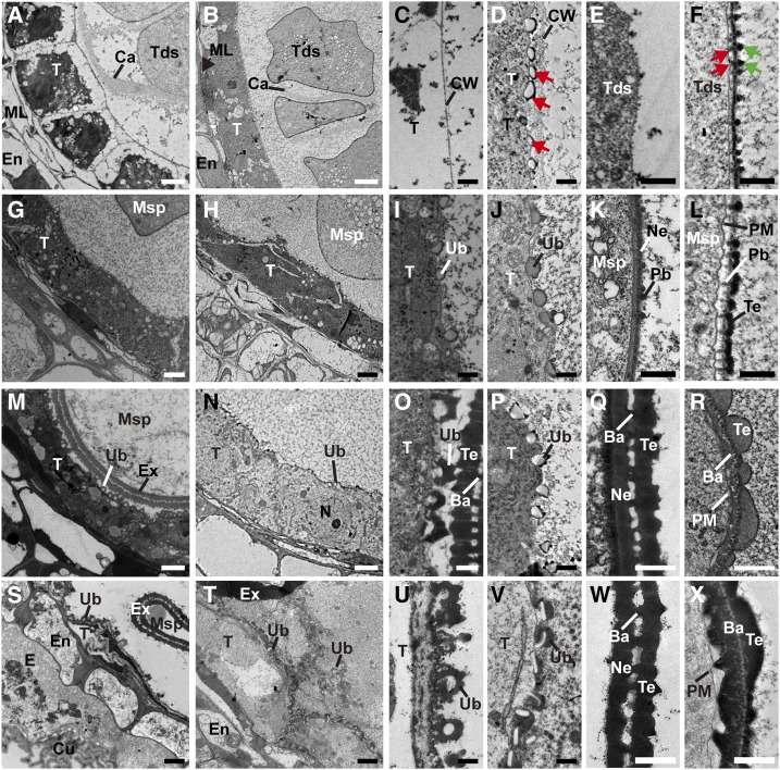
Figure 3.
TEM images of the anthers from the wild-type (WT) a and ptc2-1 mutant. Transverse sections showing tapetal cells (A and B), tapetal surface (C and D), and tetrad surface (E and F) of wild-type (A, C, and E) and ptc2-1 (B, D, and F) at stage 8b. Tapetal cells (G and H), tapetal surface (I and J), and exine (K and L) of wild-type (G, I, and K) and ptc2-1 (H, J, and L) at stage 9. Tapetal cells (M and N), tapetal surface (O and P), and exine (Q and R) of wild-type (M, O, and Q), and ptc2-1 (N, P, and R) at stage 10. Tapetal cells (S and T), tapetal surface (U and V), and exine (W and X) of wild-type (S, U, and W) and ptc2-1 (T, V, and X) at stage 11. Ba, bacula; Bu, bubble; Ca, callose; Cu, cuticle; CW, cell wall; E, epidermis; En, Endothecium; Ex, exine; ML, middle layer; Msp, microspore; N, nucleus; Ne, nexine; Pb, probacula; T, tapetum; Tds, tetrads; Te, tectum; Ub, Ubisch body. Red arrows in (D) show the bubble structure; green arrows in (F) show dot-like structure on tetrad surface and the red arrows in (F) shows a thin rod between PM and the thick layer. Scale bars = 2 μm (A, B, G, H, M, N, S, and T), 0.5 μm (C–F, I–L, O, P, U, and V), and 1 μm (Q, R, W, and X).

