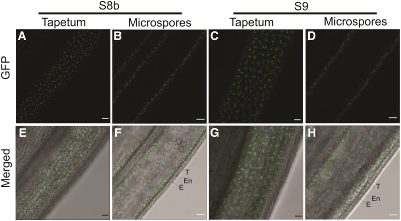
Figure 7.
Wild-type PTC2 protein localization in ptc2-1 anther. Images were acquired through a GFP filter (A–D) and merged with bright field imaging (E–H). A, B, E, and F, Anther view focusing on tapetum (A and E) and microspores (B and F) at stage 8b. C, D, G, and H, Anther view focusing on tapetum (C and G) and microspores (D and H) at stage 9. Dotted circle (F and H) showing tetrads and microspores respectively. E, Epidermis; En, endothecium; T, tapetum. Scale bars = 20 μm.

