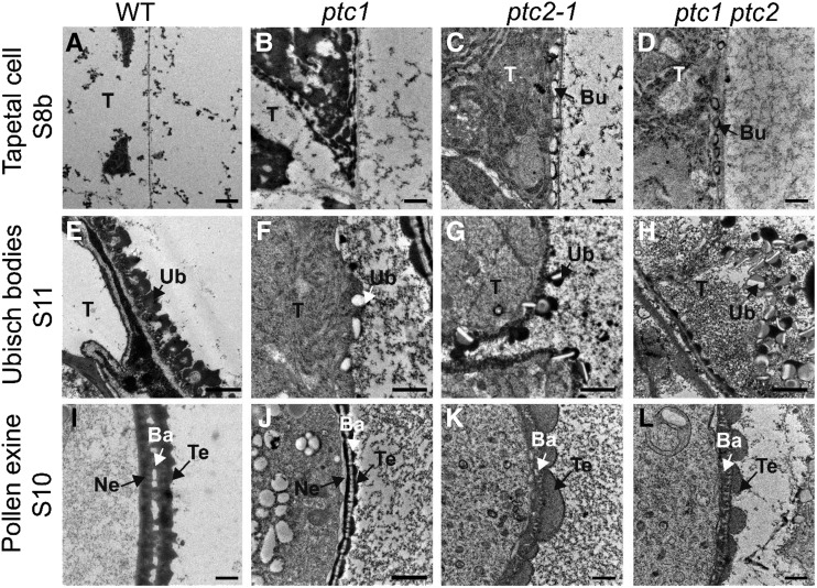
Figure 8.
TEM images of the anthers from the wild-type (WT), ptc1, ptc2-1, and ptc1 ptc2 mutants. A to D, Transverse sections showing tapetal cells at stage 8b. E to H, Transverse sections showing Ubisch bodies at stage 11. I to L, Transverse sections showing exine at stage 10. Bu, bubble-like structure; Ba, bacula; Ne, nexine; T, tapetum; Te, tectum; Ub, Ubisch body. Scale bars = 0.5 μm (A–H and J) and 1 μm (I, K, and L).

