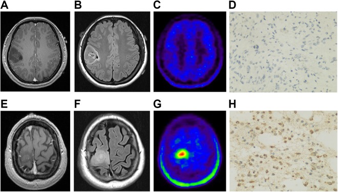
Figure 3.
Representative cases. A, T1-weighted MRI shows a low-intensity lesion in the right frontal lobe. B, Fluid-attenuated inversion-recovery MRI outlines the margin of the lesion. C, 11C-methionine PET shows weak accumulation in the lesion with SUVmax of 1.25 and TBRmean of 0.77. D, Surgery confirms the diagnosis of IDH1 mutated astrocytoma was confirmed. E, T1-weighted MRI shows a low-intensity lesion in the right frontal lobe. F, Fluid-attenuated inversion-recovery MRI outlines the margin of the lesion. G, 11C-MET PET shows strong accumulation in the lesion, with SUVmax of 8.45 and TBRmean of 3.25. H, Surgery confirms the diagnosis of IDH1 wild-type anaplastic astrocytoma was confirmed. 11C-MET PET indicates 11C-methionine positron emission tomography; IDH1, isocitrate dehydrogenase 1; MRI, magnetic resonance imaging; SUVmax, maximum standardized uptake; TBRmean, mean tumor-to-background ratio.

