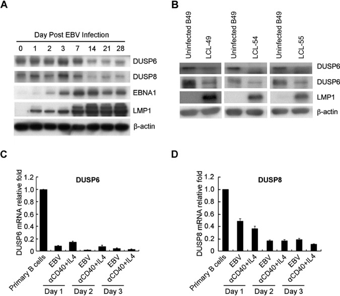
FIG 2.
DUSP6 and DUSP8 depletion caused by EBV infection. (A) Cells were infected with B95.8 strain EBV for 28 days to establish LCLs. Cell lysates of infected and uninfected B cells were analyzed for DUSP6, DUSP8, and LMP1 expression by Western blotting. β-Actin was the internal control. (B) B lymphocytes were infected with strain B95.8 EBV and harvested on the day postinfection indicated. Immunoblots of DUSP6, DUSP8, EBNA1, and LMP1 are shown. β-Actin served as an internal control. (C and D) B cells were infected with EBV or treated with anti-CD40/IL-4 for 3 days. Total RNA was extracted each day and subjected to RT-Q-PCR to detect transcripts of (C) DUSP6 and (D) DUSP8. The relative expression levels were compared to those of untreated B lymphocytes after normalization with GAPDH expression.

