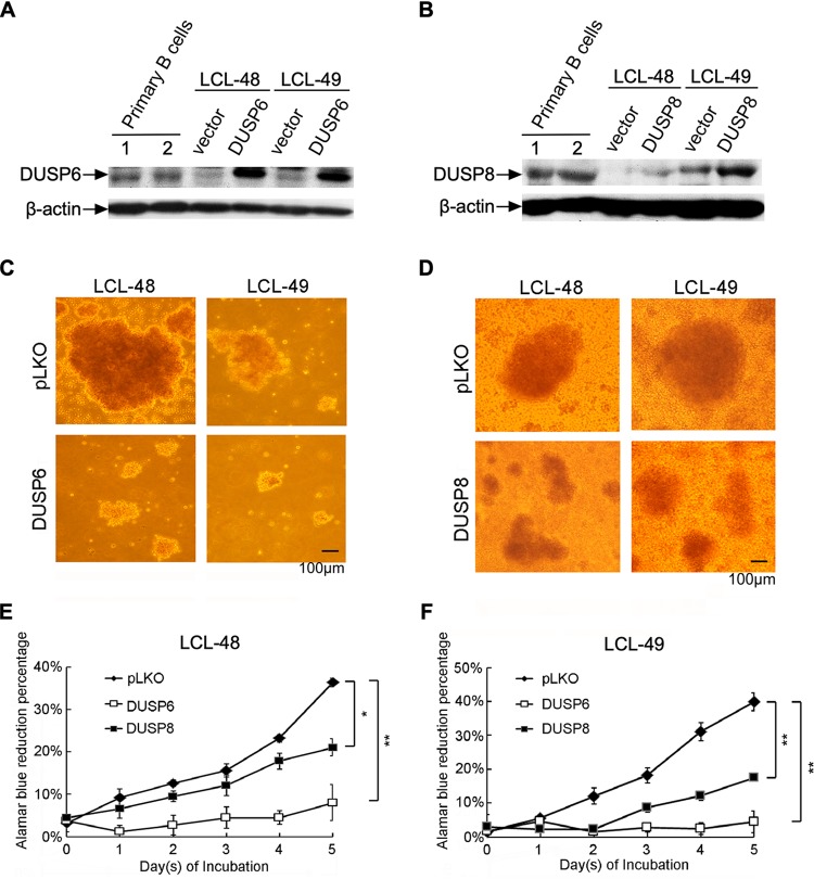
FIG 5.
Overexpression of DUSP6 and DUSP8 impaired the proliferation of LCLs. DUSP6 and DUSP8 were transduced into LCLs through lentiviral infection, and the cells were selected with G418 for 5 days. (A and B) Primary B cells or selected cells were analyzed by Western blotting to detect the indicated protein expression levels. (C and D) Selected cells were reseeded in 96-well plates at a density of 1 × 10⁶ cells per ml. Photographs were taken under a bright-field microscope after 1 day of incubation. (E and F) Selected cells were reseeded in 96-well plates at a density of 1 × 10⁵ cells per ml and incubated for 5 days. Proliferation rates of LCLs were determined by serial measurement of viable cell number every day via alamarBlue assay. (*, P<0.05; **, P<0.01 [Student's t test].)

