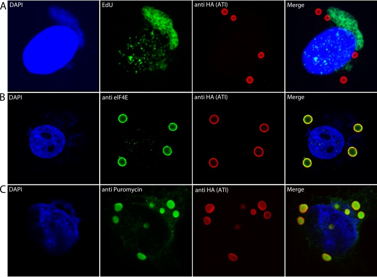
FIG 2.
Labeling of viral DNA in factories and translation of mRNA at inclusion bodies. (A) Labeling of viral DNA. HeLa cells were infected with vATI-HA.ΔA26 and incubated with EdU from 2 to 8 h. The cells were then fixed, permeabilized, and reacted with Alexa Fluor 488 azide. The cells were also stained with anti-HA antibody and a fluorescent secondary antibody to detect the ATI protein and DAPI to label nuclear and cytoplasmic viral DNA. (B) HeLa cells were infected as in Fig. 1 and incubated with mouse monoclonal anti-eIF4E and rabbit polyclonal anti-HA antibodies followed by fluorescent secondary antibodies and DAPI. (C) HeLa cells were infected as above and incubated with puromycin and cycloheximide for 30 min. After digitonin extraction and fixation, the cells were incubated with mouse monoclonal anti-puromycin and rabbit polyclonal HA antibodies followed by fluorescent secondary antibodies and DAPI. Maximum intensity projections are shown in panel A, and Z sections are shown in panels B and C. Representative images are shown.

