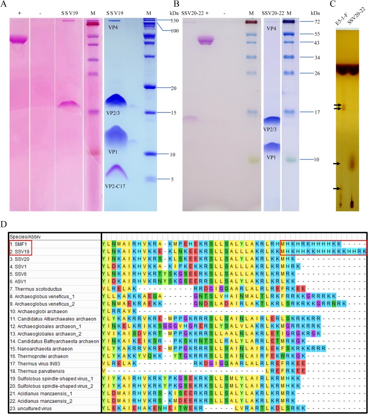
FIG 5.
Protein and lipid analyses of SSV19 and SSV20-22 virions. (A and B) Tricine–SDS-PAGE and glycoprotein staining of the virions of SSV19 (A) and SSV20-22 (B). Purified SSV20-22 virions were subjected to electrophoresis in a 14% tricine–SDS-polyacrylamide gel. The gel was stained with Coomassie brilliant blue (right) or with a glycoprotein staining kit (left). Glycosylated proteins were stained pink. +, positive control (horseradish peroxidase provided by the manufacturer); −, negative control (soybean trypsin inhibitor provided by the manufacturer); M, molecular mass markers. (C) Thin-layer chromatography of lipids extracted from SSV20-22 virions and the host cells. Differences between the lipids from SSV20-22 and those from the host cells are indicated by arrows. (D) Sequence alignment of the C-terminal regions of VP2-like proteins. Homologues of SSV19-VP2 with an e value of ≤10−3 were selected and aligned with Muscle. The C-terminal regions of SSV19 and SMF1 VP2 with similar HK repeats are shown in a red box.

