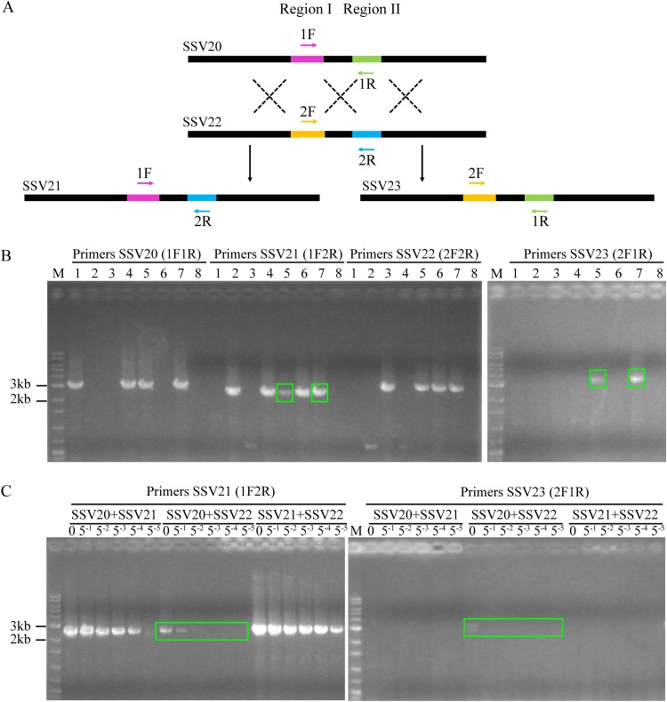
FIG 9.
Coinfection of Sulfolobus sp. strain E5-1-F by SSV20-22. (A) A diagram showing proposed crossovers between the three viruses. Primer pairs designed to detect the HR events are indicated. (B) Agarose gel electrophoresis of the products of recombination between SSV20 to SSV22. Strain E5-1 was grown to the exponential phase (OD600 of ∼0.2). The cells were infected or coinfected with different viruses at an MOI of ∼5. Lanes 1 to 8: SSV20, SSV21, SSV22, SSV20 plus SSV21, SSV20 plus SSV22, SSV21 plus SSV22, SSV20 plus SSV21 plus SSV22, and a no virus control. After incubation for 48 h, total DNA was extracted from the infected culture. PCRs were performed with indicated primer pairs using the total DNA as the template. PCR products were subjected to electrophoresis in 1.0% agarose gel. Recombination products are shown in green boxes. (C) Estimation of the frequency of recombination between SSV20 and SSV22. The total DNA extracted from the coinfected culture was serially diluted by 51- to 55-fold. PCRs targeting SSV21 and SSV23 were performed using the serially diluted total DNA as the template. Undiluted DNA is indicated by a zero; M, molecular weight standards.

