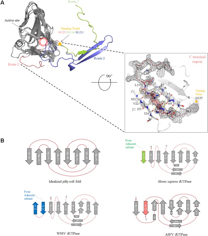
FIG 3.
(A) Three-dimensional (3D) structure and topology diagrams comparing Homo sapiens, WSSV, and ASFV dUTPases. A superposition of Homo sapiens (PDB 2HQU), WSSV (PDB 5Y5P), and ASFV dUTPase 3D structures is shown. Similar portions of the structure are shown as dark gray, and the unique C-terminal orientations are shown in salmon (ASFV), light green (Homo sapiens), and light blue (WSSV). The critical turning point is shown as a gold ball, and the active site is indicated by a pink halo (upper left). The β-turn is shown as a stick model. The C-terminal region of the ASFV dUTPase is displayed in the lower right frame, and the last β-sheet is stabilized by hydrogen-bonds with β1 and β2 strands. The electron density map (2Fo-Fc contoured at 1.0 σ) around residues after G125 is shown as a black mesh. (B) Homo sapiens, WSSV, and ASFV dUTPase have a very similar topology as the idealized jelly-roll fold.

