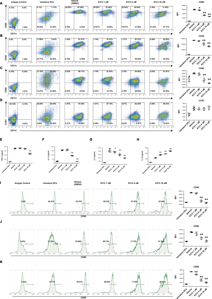
Figure 3.
K313 had the ability to suppress maturation of DCs. The sorted murine DCs were collected and treated with 1, 4, and 16 μM K313, LPS was added after 6 h. After culturing for 24 h, cells were collected and washed, then stained with indicated antibodies such as anti-murine CD11c, anti-murine CD80, anti-murine CD40, anti-murine CD86, and anti-murine CCR7, finally analyzed by flow cytometer. Flow cytometry analysis of anti-murine CD80 (A), anti-murine CD40 (B), anti-murine CD86 (C), and anti-murine CCR7 (D) expressions on murine DCs treated with K313. Histogram analysis of mean fluorescence intensity (MFI). The supernatants were gathered for ELISA test. The cytokines such as TNF-α (E), IL-12 (F), IL-6 (G), and IL-10 (H) were measured according to protocols of ELISA Kits which purchased from BD Pharmingen. Histogram analysis of each cytokine expression. Human PBMCs derived DCs were collected and treated with 1, 4, and 16 μM K313, LPS was added after 6 h. After culturing for 24 h, cells were collected and washed, then stained with indicated antibodies such as anti-human CD14, anti-human CD40, anti-human CD86, and anti-human CD80, finally analyzed by flow cytometer. Flow cytometry analysis of anti-human CD40 (I), anti-human CD86 (J) and anti-human CD80 (K) expressions on human DCs treated with K313. Histogram analysis of MFI, each column denoted the mean ± SD of six independent experiments results (*P < 0.05, **P < 0.01 versus the vehicle group).

