
Figure 1.
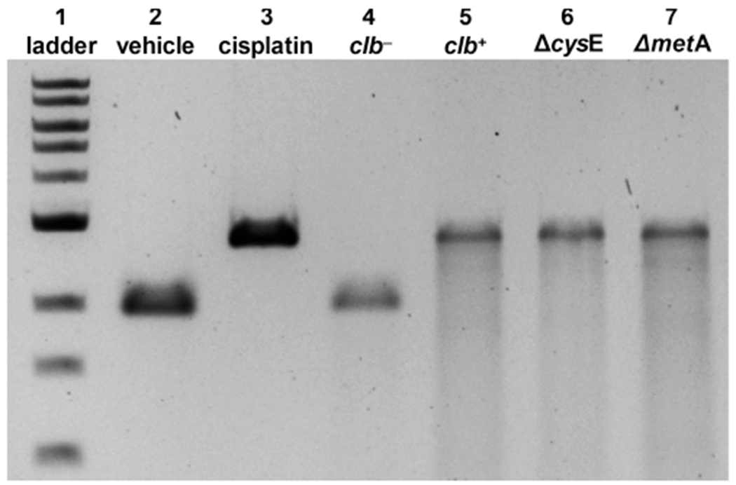
DNA cross-linking assays employing linearized pUC19 DNA and E. coli variants. Cisplatin was used as a positive control: DNA ladder (lane 1), no treatment (lane 2), cisplatin (100 μM, lane 3), clb− BW25113 E. coli (lane 4), clb+ BW25113 E. coli (lane 5), ΔcysE clb+ BW25113 E. coli (lane 6), and ΔmetA clb+ BW25113 E. coli (lane 7). Conditions for lanes 2 and 3: linearized pUC19 DNA (31 μM in base pairs), pH 5 sodium citrate buffer (10 mM), 4.5 h, 37 °C. Conditions for lanes 4 and 5: linearized pUC19 DNA, M9 medium, 4.5 h, 37 °C. Conditions for lanes 6 and 7: linearized pUC19 DNA, modified M9 medium (containing l-[U-13C]Cys or l-[U-13C]Met for Cys and Met auxotrophs, respectively), 4.5 h, 37 °C. DNA was isolated and analyzed by denaturing agarose gel electrophoresis (90 V, 1.5 h).