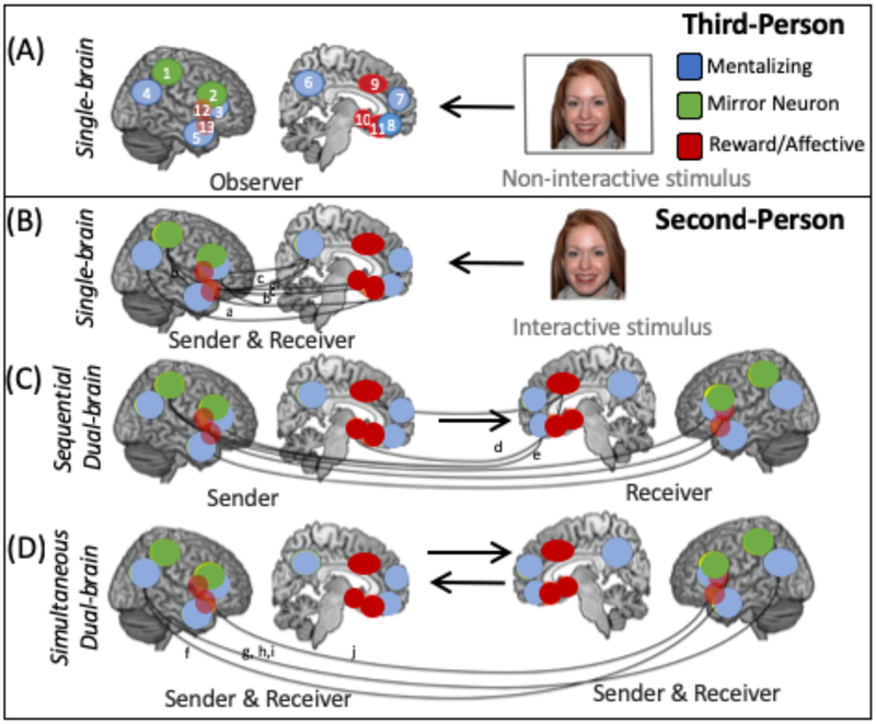
Fig. 1. Single and dual brain approaches in second-person neuroscience.
The figure depicts the different approaches that are used in second-person neuroscience studies of social cognition and compares them to third-person studies. In each panel the brain image represents the brain activity that is measured from a participant (either one or two brains are shown depending on whether a single or dual-brain approach, respectively). a| In third-person approaches, a single brain is imaged while the participant, in the role of ‘observer,’ passively perceives a non-interactive stimulus, such as a picture of a face or a video of a hand action. These approaches have highlighted regions that are part of distinct ‘social’ networks, including the mentalizing, mirror neuron and reward and/or affective networks as being important for social processing129. Several regions that are key parts of these networks are depicted. b-d| Several second-person approaches are illustrated. In each panel, the role of the participant (as the sender or receiver of communication) in the interaction is listed above the brain. These second-person methods can include single-brain approaches (b) that allow for reciprocal interaction between the participant and the interactive stimulus but in which only the brain response of the participant is examined. Examples of such an interactive stimulus include a gaze-contingent avatar, a live social partner transmitted via real-time video link or in the scanner room, or a video recording of a person directing communicative gestures towards the participant. Solid lines between brain regions depict correlations (indicating functional connectivity) within and between regions of different social networks during the sending or receiving of messages or communicative signals to an interactive stimulus20,82,97. Dual-brain second-person approaches include both sequential and simultaneous dual-brain methods. In sequential dual-brain methods (c), the timecourse and pattern of neural activity in the sender’s brain can be compared with that of the receiver to determine how one partner’s brain activity either influences the other’s brain activity unidirectionally, through the actions that it generates, or is similar to the other’s during the sending and receiving of messages. In these studies, the ‘message’ (which may be facial displays of emotion or gestures during a charade game, for example) is first recorded by the sender while their brain activity is imaged and then presented to the receiver while their brain is imaged at a later time (depicted by a unidirectional arrow). Dashed lines represent regions in which brain activity of the sender is thought to influence or be similar to that of the receiver6,95. In simultaneous dual-brain approaches (d) coupling (synchronous activity within similar brain regions in all participants) and inter-brain connectivity (correlations between neural activity in one region in one participant and neural activity in distinct regions in another participant) in the brains of participants engaged in direct social interaction can be measured. Examples of this coupling and inter-brain connectivity are depicted by dashed lines between brain images9–12,73. These single- and dual-brain approaches have demonstrated simultaneous engagement and interactions among nodes of supposedly distinct networks (that is, the mentalizing, mirror neuron, and reward networks20,28,30,60) which we propose forms an integrated social interaction network. To fully map the organization and function of this social interaction network will require further innovative methodological advances, including more ecologically-valid experimental tasks as well as quantitative measures of collective behavior and a greater focus on the emergence, development, and plasticity of this social interaction network. ACC, anterior cingulate cortex; AI, anterior insula; ATL, anterior temporal lobe; dmPFC, dorsomedial prefrontal cortex; IFG, inferior frontal gyrus; IPL, inferior parietal lobe; IPS, intraparietal sulcus; OFC, orbitofrontal cortex; PCUN, precuneus; STS, superior temporal sulcus; TPJ, temporoparietal junction; vmPFC, ventral medial prefrontal cortex; vPMC, ventral premotor cortex. VS, ventral striatum.

