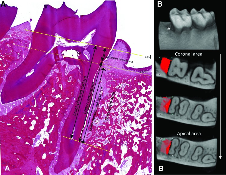
FIG. 2.
Representative micrograph indicating the scoring systems: (A) histological measurement and (B) CT measurement. (A) The landmarks for the histomorphometric measurements, the a.b.h. and c.e.j., are marked with straight dashed lines. For the principal measurements, length of root planed surface (Lroot-planed surface), length of epithelial downgrowth (Lepithelium downgrowth), length of regenerated cementum (Lnew cementum), and length of regenerated PDL (Lnew periodontal ligament), are represented with arrows. (B) The three-dimensional reconstruction of one sample with representative two-dimensional cross-sections of the reconstructed CT images from corona to apex of the defect area. *The defect area. The red area, the region of interest (defect area). a.b.h., alveolar bone height; c.e.j., cementum-enamel junction; PDL, periodontal ligament. Color images are available online.

