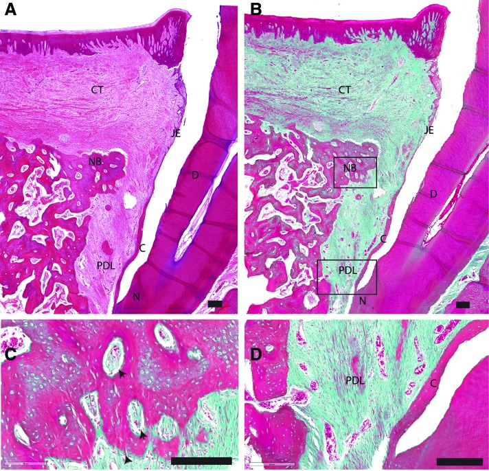
FIG. 3.
Representative overview of histological sections, H&E/trichrome stained, of the control group. Scale bar is 200 μm. (A) Lower magnification of the defect area stained in H&E. (B) Lower magnification of the defect area stained in Masson's trichrome. (C) Higher magnification of the framed area (NB) in (B). (D) Higher magnification of the framed area (PDL) in (B). black arrow: osteoids with osteoblasts. C, cementum; CT, connective tissue; D, root dentin; H&E, hematoxylin and eosin; JE, junctional epithelium; N, notch of planed surface; NB, new bone; R, gingival epithelium. Color images are available online.

