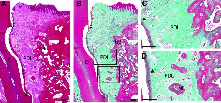
FIG. 4.
Representative overview of histological sections, H&E/trichrome stained, of the experimental group. Scale bar is 200 μm. (A) Lower magnification of the defect area stained in H&E. (B) Lower magnification of the defect area stained in Masson's trichrome. (C) High magnification of the framed area (upper) in (B). (D) High magnification of the framed area (lower) in (B). black arrow: PDL inserted into new formed cementum. Color images are available online.

