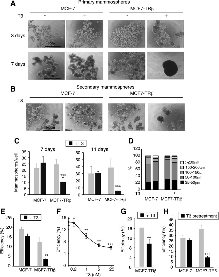
FIG. 1.
T3 inhibits mammosphere formation and expression of pluripotency markers in TRβ-expressing MCF-7 cells. (A) Representative optical microscope images of MCF7-TRβ and MCF-7 cells growing under nonadherent conditions for three and seven days in the presence and absence of 25 nM T3. (B) Images of 11-day second-generation mammospheres obtained after dispersion of 3-day primary mammospheres. Scale bars = 100 μM. (C) Quantification of 7- and 11-day secondary mammospheres grown with and without T3. (D) Size distribution of seven-day secondary mammospheres. (E) Cells were plated at a limiting dilution in 96-well plates under nonadherent conditions, and the efficiency of mammosphere formation was scored after 14 days in the presence and absence of T3. (F) MCF7-TRβ cells were inoculated at a limiting dilution and grown under nonadherent conditions in the presence of the indicated concentrations of T3. The number of mammospheres generated was scored after 14 days. Data are mean ± SD of three independent 96-well plates, and significance with respect to the untreated cells is shown with asterisks. (G) MCF7-TRβ cells were grown as primary mammospheres for three days with and without T3, dispersed, and plated at a limiting dilution in 96-well plates. The efficiency of mammosphere formation is shown. (H) Adherent 6-well cultures were incubated with and without T3 for 10 days, dispersed, inoculated at a limiting dilution in 96-well plates, and grown under nonadherent conditions. Mammospheres were counted after 14 days of incubation in the absence of T3. Data are mean ± SD of three independent plates. Asterisks denote the existence of statistically significant differences between T3-treated and untreated cells. **p < 0.01 and ***p < 0.001. T3, triiodothyronine; TR, thyroid hormone receptor.

