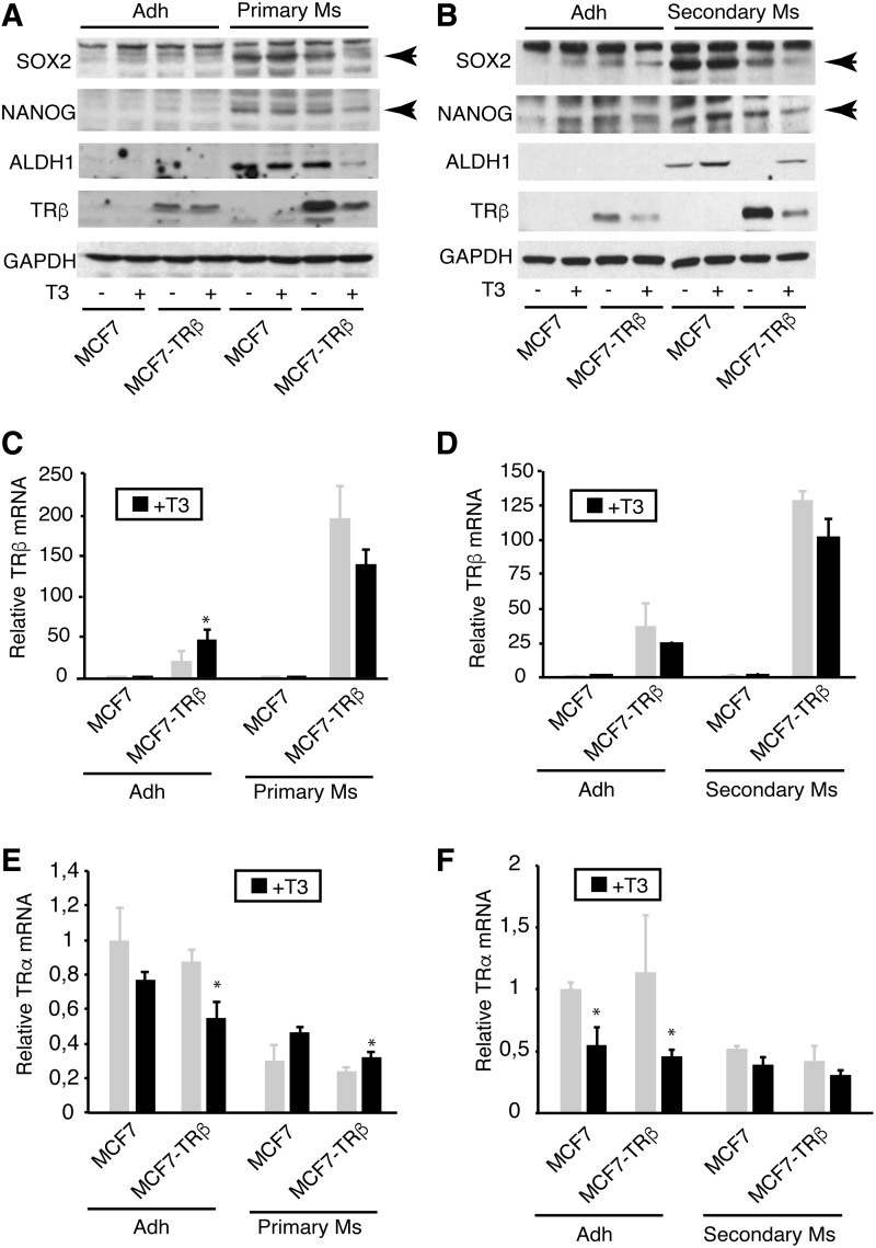
FIG. 2.
T3 inhibits expression of pluripotency markers. (A) The levels of SOX2, NANOG, ALDH1, and TRβ were determined by Western blotting in MCF-7 and MCF7-TRβ cells grown under adherent conditions (adh) or as primary mammospheres (Ms) for three days in the presence and absence of T3. GAPDH was used as a loading control. (B) Similar experiment performed in adherent cultures and seven-day secondary mammospheres derived from three-day primary mammospheres grown with and without T3. Arrows indicate the specific bands. (C) TRβ mRNA levels in untreated and T3-treated adherent cultures and in primary mammospheres. (D) TRβ mRNA levels in adherent cultures and secondary mammospheres incubated in the presence and absence of hormone. (E) TRα mRNA levels in untreated and T3-treated adherent cultures and primary mammospheres. (F) TRα transcripts in secondary mammospheres and adherent cells treated with and without T3. Data are mean ± SD. Statistically significant differences between T3-treated and untreated cells are shown by asterisks. *p < 0.05. ALDH1, aldehyde dehydrogenase 1; GAPDH, glyceraldehyde 3-phophate dehydrogenase; NANOG; SOX2, sex determining region Y-box 2.

