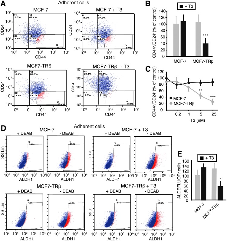
FIG. 3.
TRβ mediates a T3-dependent decrease of the CD44+CD24− and ALDH+ cell subpopulations. (A) Flow cytometry analysis of the expression of CD44 and CD24 in adherent cultures of parental and TRβ-expressing MCF-7 cells treated with or without 25 nM T3 for 72 h. The threshold lines were set according to the isotype control. Gating of the more extreme CD44+CD24− cell population shows its disappearance in T3-treated MCF7-TRβ cells. (B) Quantification of the CD44+CD24− cell population obtained in four independent experiments. Data are expressed as the percentage of the value obtained in the parental control cells without T3 treatment and are mean ± SD. (C) Quantification of CD44+CD24− cells after 72-h incubation of MCF-7 and MCF7-TRβ cells, with the concentrations of T3 indicated. Data (mean ± SD) were obtained from two independent experiments performed in duplicate and are shown as the percentage of the results obtained in the untreated MCF-7 cells. Significant differences between MCF-7 and MCF7-TRβ cells are shown with asterisks. (D) ALDH enzymatic activity as assessed by ALDEFLUOR assays and flow cytometry in adherent cultures of parental and TRβ-expressing MCF-7 cells incubated in the presence and absence of 25 nM T3 for 72 h. To determine the percentage of ALDEFLUOR+ cells, assays were performed in the presence and absence of the ALDH-specific inhibitor DEAB. (E) Relative percentage of ALDEFLUOR+ cells (mean ± SD) obtained from three independent experiments. Significant differences between T3-treated and the corresponding untreated cells are shown with asterisks. *p < 0.05, **p < 0.01, and ***p < 0.001. DEAB, diethylaminobenzaldehyde.

