Fig. 3.
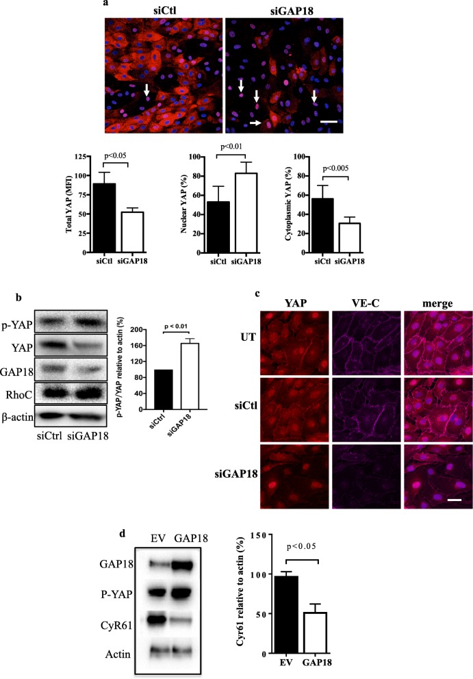
ARHGAP18 depletion results in YAP activation. a Cells were treated with siCtl or siARHGAP18 (siGAP18) and placed under 48 h of laminar flow. Representative images showing nuclear YAP (pink, white arrows) in ECs lacking ARHGAP18 under high shear flow. Nuclei (blue). Scale bar = 50 μm. Quantification of MFI for n = 3 experiments of total, nuclear and cytoplasmic YAP. b Cells were depleted of ARHGAP18 by siRNA and then total YAP or p-YAP measured 72 h later. A representative western blot and quantification of n = 3 experiments is shown. c ECs were untreated (UT), treated with siCtl or siARHGAP18 (siGAP18) and 48 later imaged for YAP and VE-cadherin expression. c Cyr61 protein expression in ECs lysates 6 days after EV or ARHGAP18 (GAP18) infection. β-actin was used as a loading control. Quantification of densitometric ratio for Cyr61relative to actin. Data are presented as the normalised expression of mean of 3 independent EC lines ± SD. p by paired t-test

