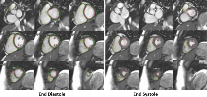
Fig. 2.
Contours of the right ventricle (RV) and left ventricle (LV) at end-diastole (left panel) and end-systole (right panel) demonstrating contouring strategy. Papillary muscle and trabeculations are included in the blood pool by tracing the blood pool-endocardial border of each ventricle in both phases

