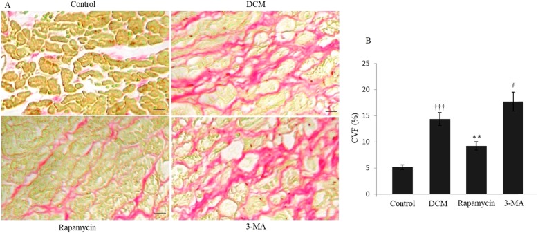
Fig. 1.
Modulating autophagy and cardiac matrix remodeling of DCM. (A) Picrosirius red staining indicated significantly changes of collagen distribution in the four different groups. (B) Histochemical analysis showed that there was a significant increase of collagen distribution in the DCM group compared with the control group. Quantitative assessment demonstrated that the CVF was significantly decreased in the rapamycin group, and it was increased in the 3-MA group compared with the DCM group. †††P < 0.001 vs Control, **P < 0.01 and #P < 0.05 vs DCM. Scale bar = 100 μm

