Figure 5.

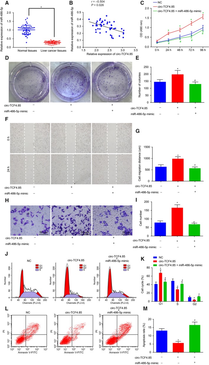
MicroRNA‐486‐5p overexpression disrupts the promoted HCC cell viability, colony‐forming ability, migration, and invasion induced by circ‐TCF4.85. HCC cells were transfected with miR‐486‐5p mimic in the presence of circ‐TCF4.85. (A) MicroRNA‐486‐5p expression patterns in HCC tissues and adjacent normal tissues detected by RT‐qPCR. (B) Correlation analysis of circ‐TCF4.85 and miR‐486‐5p. (C) HCC cell viability in each group detected using MTT assay. (D) HCC cell colony formation ability in each group detected using clonogenic assay. (E) Colony‐forming cell number in each group. (F) HCC cell migration in each group detected using scratch test. (G) Migration distance in each group. (H) HCC cell invasion in each group detected using Transwell assay (×400). (I) Invasive cell number in each group. (J) Cell cycle progression detected by flow cytometry. (K) Proportion of cells at G1, S, and G2 phases in each group. (L) Cell apoptosis in each group detected by flow cytometry. (M) Apoptotic rate of HCC cells in each group. *P < 0.05, compared with the NC group; # P < 0.05, compared with the circ‐TCF4.85 group. Data were expressed as mean ± SD. Comparisons between adjacent normal tissues and HCC tissues were analyzed using t‐test (n = 46), and those among multiple groups were analyzed by the one‐way ANOVA or repeated‐measures ANOVA. Correlation analysis between two groups was conducted using Pearson’s correlation analysis. The experiment was repeated three times.
