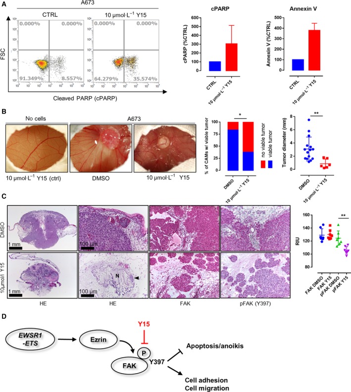
Figure 3.

Effects of Y15 treatment on EwS viability and invasion in an in vivo (avian CAM) model. (A) FACS analyses confirmed the proapoptotic effect of FAK inhibition on A673 EwS cells. (B) Application of 10 µm Y15 to A673 EwS cells 24h prior to cell seeding significantly impaired the rate of tumor formation as well as tumor size in the CAM model (P = 0.0122 and P = 0.0095, respectively; n = 34 and n = 21, Fisher’s exact test and unpaired t‐test) while not altering the (micro)vascular architecture of the CAM. (C) Histological analyses showed tumor regression, necrosis (N), and microcalcification with only very few residual tumor cells detectable (arrowhead) upon Y15 treatment. Immunohistochemistry and quantification via reciprocal intensity showed persistent expression of FAK in both Y15‐treated and control cells, but a significant decrease in FAK Y397 phosphorylation in the residual A673 xenograft tumor cells after treatment with Y15 (n = 21, Sidak’s multiple comparison test). (D) Schematic diagram of how EWSR1‐FLI1‐dependent expression of Ezrin may contribute to SRC‐independent autophosphorylation of FAK on tyrosine 397 as previously described. The phosphorylation of FAK impairs apoptosis and anoikis and enhances focal adhesion formation and migratory capacity of EwS cell, but can effectively be targeted by application of Y15. *p < 0.05; **p < 0.01; ***p < 0.001.
