
Figure 1.
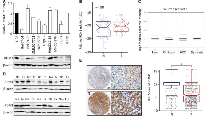
RDM1 expression is decreased in HCC. (A) The mRNA and protein expression of RDM1 in HCC cell lines and immortalized liver cell L‐02 were examined by qRT‐PCR and western blot. Statistical data were represented as mean ± SD. (B) qRT‐PCR determined RDM1 mRNA in 50 paired HCC fresh tissues and analyzed by paired t‐test. N, nontumor; T, tumor. (C) The expression of RDM1 mRNA was shown in Oncomine Wurmbach dataset. (D) RDM1 protein levels in 10 paired HCC and adjacent nontumor fresh tissues were shown by western blot. β‐actin was used as the indicator of the amount of loading proteins. (E) The expression of RDM1 in 755 paraffin‐embedded specimens was determined by TMA‐based IHC staining. The representative images of tumor (T) and nontumor (N) were presented, and the IHC score of each case was shown. The length of scale bars was 100 μm (left) and 20 μm (right). *P < 0.05.