
Figure 5.
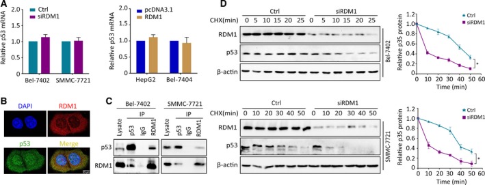
RDM1 post‐transcriptionally stabilizes p53 protein. (A) With the silencing or overexpression of RDM1, p53 mRNA levels were detected by qRT‐PCR. (B) IF staining indicating the colocalization of p53 (green) and RDM1 (red) together with DAPI (blue) in HepG2 cells. The length of scale bars was 10 μm. (C) Co‐IP assays were performed to determine the interaction of RDM1 and p53. (D) The half‐life of p53 protein was detected in RDM1‐depleted cells supplying with CHX (20 μg·mL−1) at different times. The total amount of p53 protein was quantitated and calculated by ImageJ software. All the experiments were done in triplicate. Student’s t‐test was used to analyze the statistical difference. *P < 0.05.