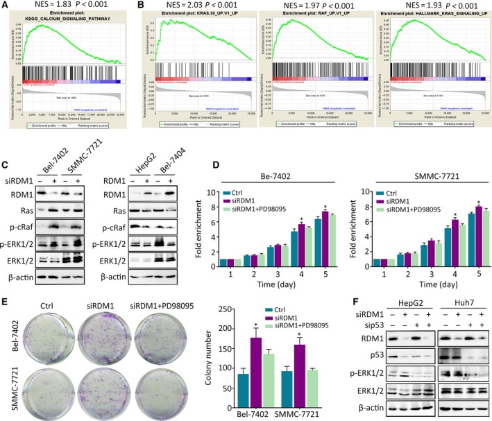
Figure 6.

RDM1 represses Ras/Raf/ERK signaling in the presence of p53. (A, B) GSEA of HCC dataset showed the correlated of RDM1 expression obtained from TCGA with related pathways. (C) Western blot validated the differential expression of Ras/Raf/ERK signaling pathway in RDM1 overexpression or depleted cells. (D, E) MTT assay (D) and colony formation assays (E) indicated the cell growth in RDM1‐depleted or ERK inhibitor PD98095‐treated groups. (F) Western blot detected the expression of p‐ERK with the depletion of p53 and RDM1 in HepG2 and Huh7 cells. All the experiments were done in triplicate. One‐way ANOVA method was used to analyze the statistical difference. *P < 0.05.
