Figure 6.

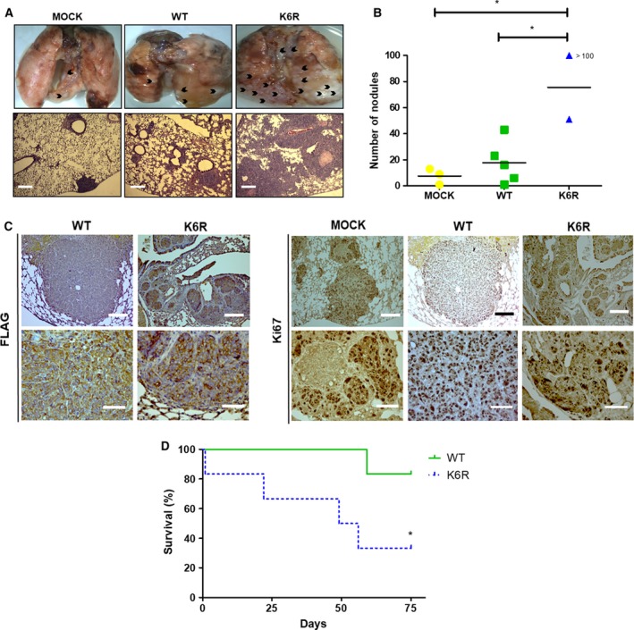
Stem‐like ECE1cK6R cells display enhanced lung colonization capacity in vivo. (A) DLD‐1 cells expressing either Flag‐tagged ECE1cWT or ECE1cK6R proteins (2 × 10⁶) were intravenously injected into NOD/SCID mice. Animals were euthanized after 75 days of monitoring, or earlier if the animals showed signs of suffering. Representative images of metastatic lungs are shown, where black arrowheads indicate metastatic nodules (upper panel), along with HE staining of paraffin sections (lower panel; scale bar = 300 µm) (B) Numbers of metastatic nodules in each cohort from mice in A were determined from paraffin slices. (C) Paraffin slices of lungs from experiment in A were analyzed using IHC with anti‐FLAG (left) and anti‐Ki‐67 (right) antibodies (scale bars upper panels = 150 µm; lower panels = 50 µm). (D) Survival (%) of mice from experiment in A. Data represent average ± SEM (n = 3 each cohort). ANOVA and Tukey tests were used, except for B: log‐rank test. *P ≤ 0.05.
