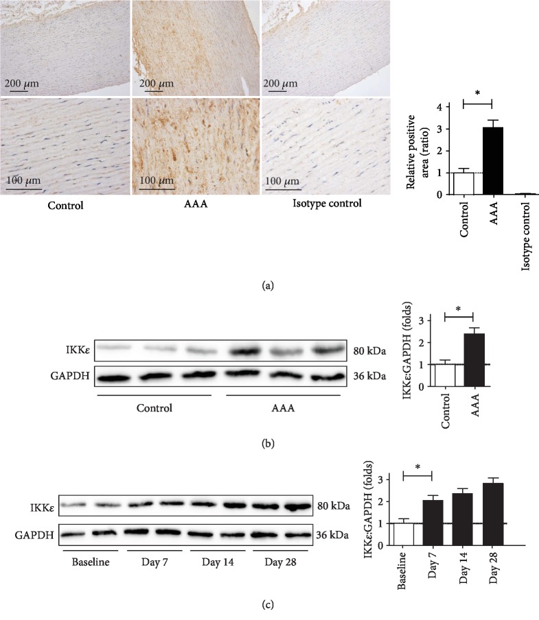
Figure 1.
The expression of IKKε is increased in the aortic aneurysm. (a) Representative microscopic photos of immunohistochemical staining for IKKε expression in the control and AAAs. (b) Immunoblots to analyze the expression of IKKε in control human nonaneurysmal aortas vs. AAAs, n = 4‐5, respectively. (c) Time course of IKKε expression after infusion with Ang II for 7, 14, and 28 days. Western blot analysis of IKKε protein level in different groups, n = 4‐5, respectively.

