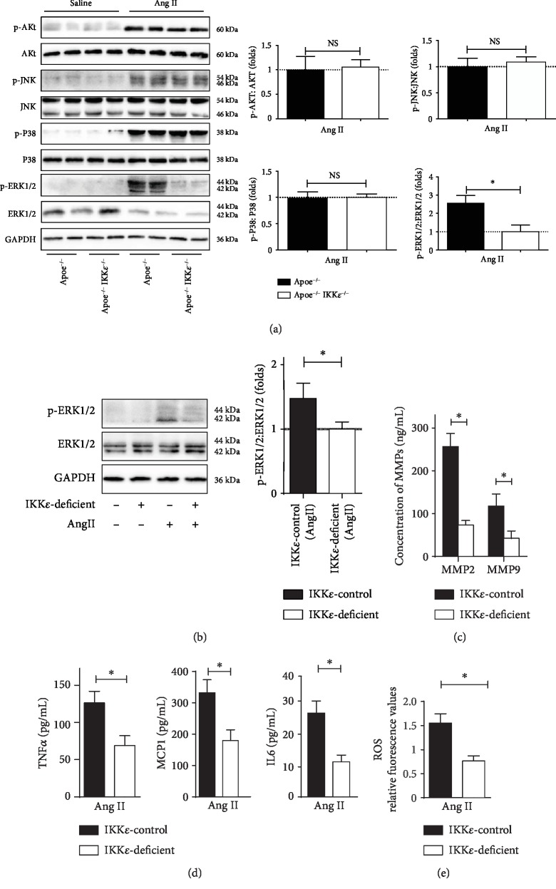
Figure 6.
IKKε deficiency blocked phosphorylation of ERK1/2. (a) Western blot analysis for the total and phosphorylated protein levels of AKT, JNK, P38, and ERK1/2 in aortic tissues from mice in the indicated groups at 28 days of infusion. Quantification shown in the right panels, n = 4‐5, respectively. ∗P < 0.05 vs. Apoe−/−IKKε−/− mice. (b) Total and phosphorylated protein levels of ERK1/2 in primary VSMC in the indicated groups. Quantification shown in the right panels. (c) MMP expression in primary VSMC in the indicated groups detected by ELISA. (d) TNFα, MCP1, and IL6 expressions in primary VSMC in the indicated groups detected by ELISA. (e) Analysis of ROS generation in primary VSMC in the indicated groups by flow cytometer, n = 4‐5, respectively.∗P < 0.05 vs. IKKε-deficient.

