
Figure 1.
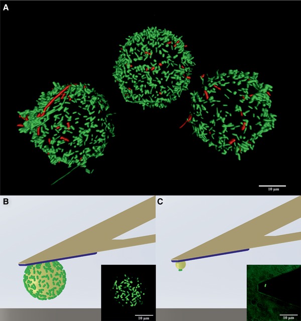
(A) 3D fluorescence image of E. coli‐PEI‐beads (Ø 20 μm) stored for 6 h in PBS. Living cells appear green, dead cells appear red.
(B and C) Schematic illustrations and corresponding fluorescence images of cell probes: (B) with a monolayer of E. coli, bead diameter 20 μm; (C) with a single E. coli, bead diameter 5 μm (according to 28).