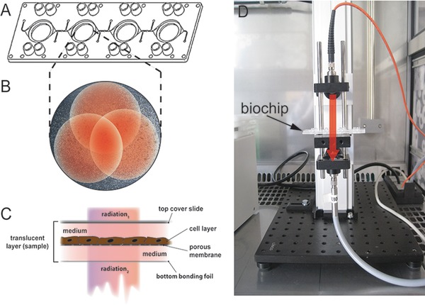
Figure 1.

(A) Multiorgan tissue flow (MOTIF) biochip with four interconnected round cavities in microscopic slide format. Each cavity comprises a porous membrane that separates the cavity in a top and a bottom chamber. (B) Example for the three partly overlapping measurement points (red dots) in one cavity with adherent HepaRG cells. (C) Schematic cross‐section of the translucent biochip containing a HepaRG cell layer and a membrane serving as cell substrate. At top, the biochip is sealed with a polystyrene cover slide. Light beam for NIRS enters the biochip through the cover slide (radiation 1) and leaves the biochip through the bottom bonding foil (radiation 2). (D) Transmission measurement setup (NIRS). The red arrow indicates the radiation pathway through the translucent biochip.
