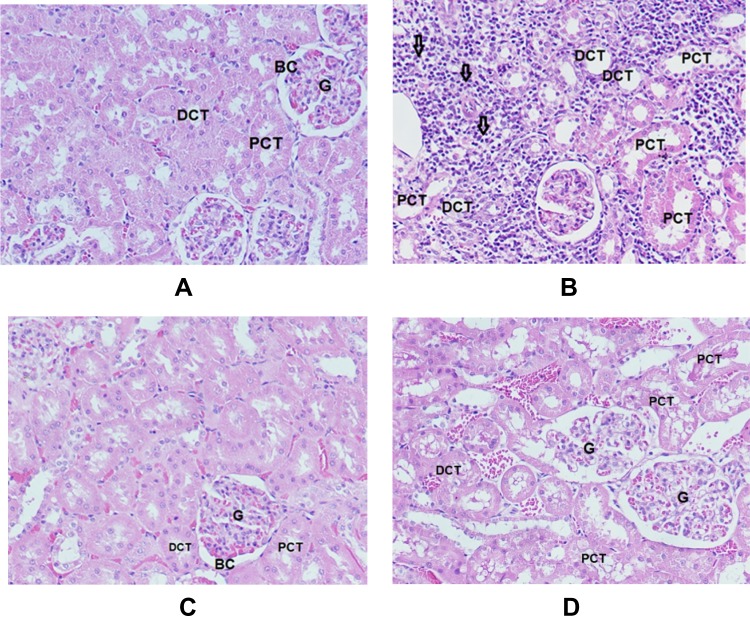
Figure 6.
Sections in cortex of the kidney (H&E x 200). (A) Normal control rats (G1) show renal corpuscle consists of glomerulus (G) and a two-layered glomerular Bowman’s capsule (BC) that encloses glomerulus; the proximal convoluted tubules (PCT) and distal convoluted tubules (DCT). (B) GNPs-treated rats (G2) show inflammatory cells infiltration, mainly in the interstitial tissue (arrows), vacuolar degeneration surrounding PCT and DCT, tubule necrosis (star), dilation and vacuolar degeneration in PCT and DCT. (C) GNPs + Vitamin E (G3) shows normal renal corpuscle surrounded by normal tubules. (D) GNPs + α-lipoic acid (G4) shows vacuolar degeneration in PCT and DCT.

