
Figure 1.
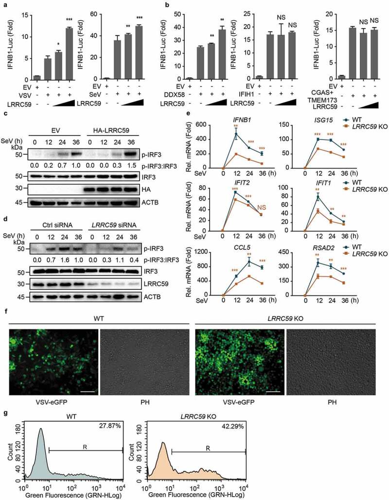
LRRC59 potentiates DDX58-mediated type I IFN antiviral immune response. (a,b) Luciferase activity in 293T cells transfected with an IFNB1 luciferase reporter (IFNB1-Luc), together with an empty vector (EV) or increasing amounts (100 ng and 300 ng per well, the same with following experiments) of plasmid encoding LRRC59, followed by infection with or without VSV or SeV (A) or activated by DDX58 (with IC poly[I:C] treatment; 5 μg/ml for 12 h in all IC poly[I:C] stimulation if no additional annotation), IFIH1 or co-expression of CGAS and TMEM173 together (CGAS+TMEM173) (b). (c) Immunoblot analysis of extracts of 293T cells transfected with EV or HA-LRRC59 and infected with SeV for the indicated time points. (d) Immunoblot analysis of extracts of A549 cells transfected with control siRNA or LRRC59-specific siRNA, followed by the infection with SeV for indicated time points. (e) Real-time PCR analysis of IFNB1 and other ISGs in A549 wild type (WT) cells or LRRC59 knockout (KO) A549 cells after SeV infection. (f,g) Phase-contrast (PH) and fluorescence microscopy analyses (f) or flow cytometric analyses (g) of WT and LRRC59 KO A549 cells infected with VSV-eGFP for 18 h. Scale bars, 200 μm. Data in (a, b, and e) are means ± SD of 3 independent experiments. *P < 0.05, **P < 0.01 and ***P < 0.001. Data in (c, d, f, and g) are representative of 3 independent experiments.