
Figure 5.
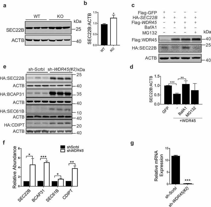
WDR45 mediates degradation of target ER proteins. (a and b) Western blot detection of endogenous SEC22B in mid-brain. Protein abundance was quantified in (b). (c and d) Co-expression of WDR45 and SEC22B in HeLa cells and protein levels were examined by western blot. Some groups of cells were treated with MG132 (20 μM, 12 h) or BafA1 (50 nM, 8 h), respectively. Protein abundances were quantified in (d). (e and f) Stability of exogenously expressed SEC22B, BCAP31, SEC61B and CDIPT in HeLa cells with WDR45 stably knocked down. Bar graph (f) shows statistics of protein expression levels. (g) Real-time PCR quantification of WDR45 mRNA in stable cell lines (#2). Data were expressed as mean ± SEM (n = 3). P values: *P < 0.05, **P < 0.01, ***P < 0.001.