Figure 6.

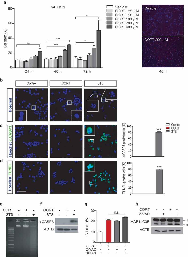
CORT treatment does not induce apoptosis or necroptosis in HCN cells. (A) Death rate of HCN cells after CORT treatment (n = 4). Right, representative image of Hoechst and PI staining 48 h after CORT treatment in HCN cells. (B) Nucleus condensation assay with Hoechst staining. (C) Immunostaining of cleaved CASP3 (c.CASP3). (D) Nuclear fragmentation assay by TUNEL staining. Scale bar: 40 μm for b-d. (E and F) Agarose gel electrophoresis of DNA fragmentation assay (E) and western blots of c.CASP3 (F) are representative of at least 3 experiments with similar results. All apoptotic markers were analyzed after CORT (200 µM for 48 h, except western blotting analysis of c.CASP3 with 72 h) or staurosporine (STS, 0.5 µM for 6 h) treatment. (G) Effects of Z-VAD (25 μM) or necrostatin-1 (NEC-1, 100 μM) on HCN cell death after CORT treatment for 48 h (n = 3). (H) Western blotting analysis of the effects of Z-VAD (25 μM) on autophagy flux after CORT treatment for 48 h. The blots are representative of 3 experiments with similar results. *P < 0.05, **P < 0.01, ***P < 0.001. n.s., not significant.
