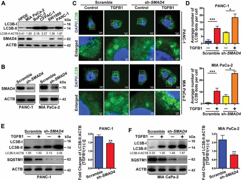
Figure 4.

SMAD4 is involved in autophagy induction by TGFB1 in SMAD4-positive PDAC cells. (A) The levels of LC3B and SMAD4 expression were examined in pancreatic cancer cell lines and HPDE cells after 5 ng/ml TGFB1 treatment for 24 h. (B) Western blotting indicated that PANC-1 and MIA PaCa-2 cells were transfected with shRNA against SMAD4 to silence SMAD4 expression. (C) The effect of TGFB1 on GFP-LC3B dot formation was evaluated in PANC-1 and MIA PaCa-2 cells with silencing of SMAD4 (Magnification scale bar, 5 μm; scale bar in enlarged image, 1 μm). (D) After PANC-1 and MIA PaCa-2 cells were SMAD4 silenced and treated with TGFB1, the number of GFP-LC3B dots per cell was quantified. (E and F) Western blot analysis showed that silencing of SMAD4 altered the effect of TGFB1 (5 ng/ml) on the conversion of LC3B-I to LC3B-II and on SQSTM1 expression in PANC-1 and MIA PaCa-2 cells. TGFB1 induced a fold change of the LC3B-II:ACTB ratio in cells with silencing of SMAD4.
