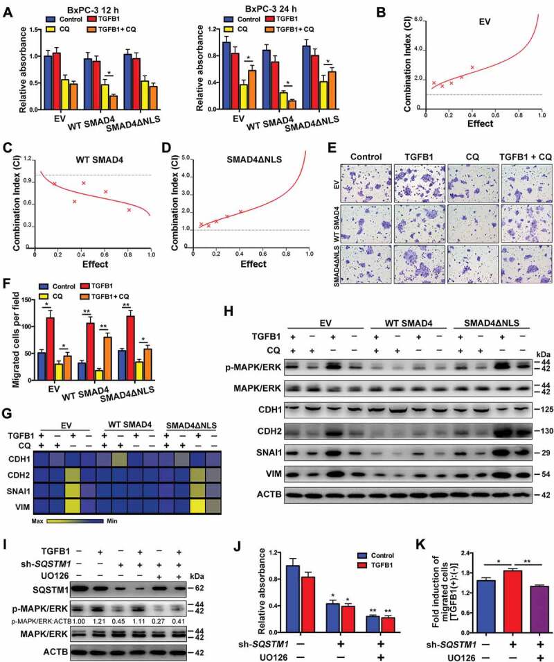
Figure 8.

Roles of TGFB1-induced autophagy in SMAD4-negative PDAC. (A) Treatment with CQ and TGFB1 for 12 h (left panel) and 24 h (right panel) affected the number of viable BxPC-3 cells transfected with the WT SMAD4 or SMAD4ΔNLS vector. (B) An antagonistic effect of CQ combined with TGFB1 was observed according to assessment of the CI of BxPC-3 cells transfected with the EV. (C) A synergistic effect of CQ combined with TGFB1 was observed according to the assessment of CI of BxPC-3 cells transfected with the WT SMAD4 vector. (D) An antagonistic effect of CQ combined with TGFB1 was observed according to the assessment of CI of cells transfected with the SMAD4ΔNLS vector. (E) Treatment with CQ and TGFB1 for 24 h affected the migration of BxPC-3 cells transfected with the WT SMAD4 or SMAD4ΔNLS vector (Original magnification, ×200). (F) The number of migrated cells per field was quantified. (G) Western blot analysis showed the effects of CQ and TGFB1 on the expression of EMT markers in BxPC-3 cells with transfection of WT SMAD4 or SMAD4ΔNLS vector. (H) Heat map showing the effects of CQ and TGFB1 on the mRNA levels of EMT markers in BxPC-3 cells with transfection of the WT SMAD4 or SMAD4ΔNLS vector. (I) Western blot analysis showed the effects of SQSTM1 knockdown on MAPK/ERK kinase activation in BxPC-3 cells. (J) The effects of SQSTM1 knockdown on the number of viable BxPC-3 cells and (K) TGFB1-induced migration were evaluated after treatment with or without TGFB1.
