
Figure 2.
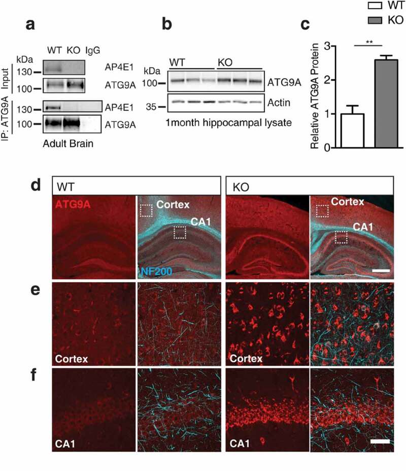
ATG9A handling is affected in vivo in ap4e1 KO mice. (a) Endogenous co-immunoprecipitation of AP4E1 with ATG9A from mouse brain, showing interaction between AP-4 and ATG9A (n = 3 independent experiments). (b) Western blot of ATG9A in hippocampus at 1 month showing increase in KO animals. (c) Quantification of relative ATG9A protein (n = 3 animals). (d) Sections prepared from AP-4 line at 1 month stained against ATG9A and NEFH/NF200, showing increased immunoreactivity and accumulation of ATG9A within neuronal cell layers. Scale bars: 200 and 50 μm (e) High magnification showing accumulation of ATG9A within cortex and (f) CA1 of hippocampus. Scale bar: 20 μm (n = 5 animals). Quantified data is expressed as mean ± SEM. Statistical analysis: Two-tailed unpaired Student’s t-test, **p < 0.01.