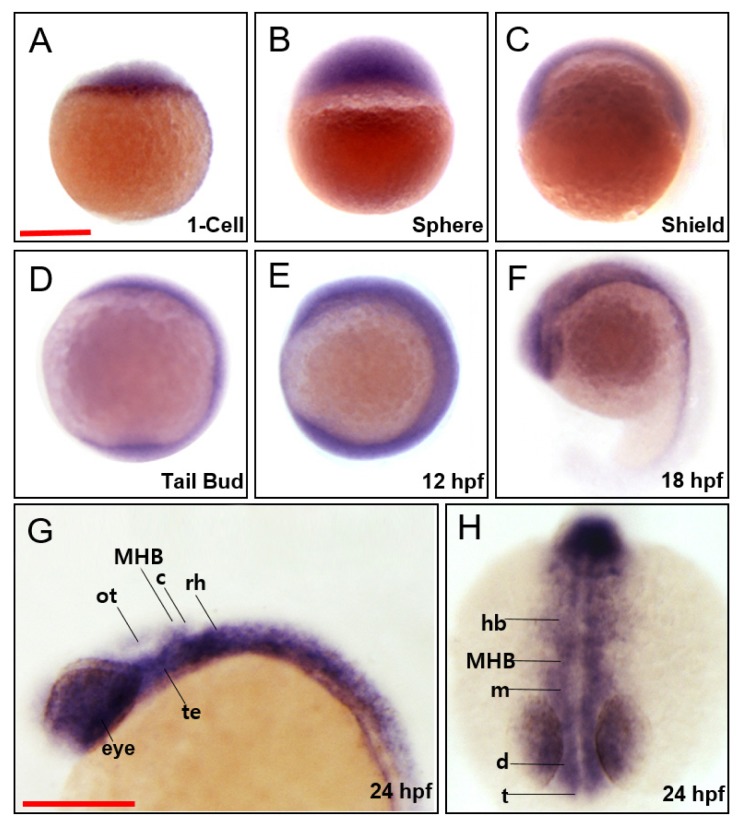
Fig. 1. Spatiotemporal expression patterns of zebrafish march5.
WISH analysis of march5 at 1 cell through 24 hpf. (A) march5 was expressed in the blastodisc at 1-cell stage, indicating it is maternally expressed. (B) After MBT (512-cell), march5 was expressed in deep cell layer (DEL) and enveloping layer (EVL) except I-YSL (yolk syncytial layer). (C) march5 was expressed in both ventral & dorsal region at shield stage. (D–F) The transcripts were abundant in the central nervous system at 10 hpf through 18 hpf. (F) At 18 somite, march5 transcripts were distributed in the precursor region of brain along the AP axis. (G and H) march5 expression patterns at 24 hpf zebrafish embryo stage. Lateral (G) and anterior view (H) of the embryos labeled with march5 antisense probe at 24 hpf. march5 was expressed in the forebrain through the notochord including the telencephalon, diencephalon, tegmentum, optic tectum, cerebellum and rhombomere. All embryos were collected synchronously from WT zebrafish for WISH analysis at the corresponding stages. MHB, midbrain hindbrain boundary; ot, optic tectum; c, cerebellum; rh, rhombomere; te, tegmentum; hb, hindbrain; m, midbrain; d, diencephalon; t, telencephalon. (A–H) Scale bars = 250 μm.

