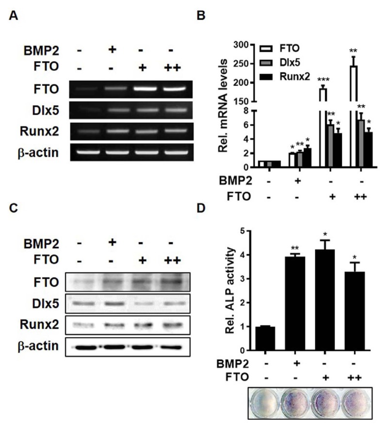
Fig. 2. Overexpression of FTO induces osteogenic differentiation of C3H10T1/2 cells.
(A–C) C3H10T1/2 cells were transfected with pcDNA3.1 (2 μg) or pCMV-FTO (+, 1 μg; ++, 2 μg) for 6 h and treated with BMP2 (0.25 μg/ml) for 1 day. (A) RT-PCR analysis was performed using total RNA isolated from cells and primers targeting FTO, Dlx5, Runx2, and β-actin. (B) Real-time PCR was performed using total RNA isolated from cells. (C) Western blot analysis was performed using the indicated antibodies. (D) C3H10T1/2 cells were transfected with pCMV-FTO (+, 0.2 μg; ++, 0.4 μg) or treated with BMP2 (0.25 μg/ml) for 4 days. *P < 0.05, **P < 0.001, and ***P < 0.005 compared with untreated control cells. Data represent the mean ± SEM of three individual experiments. All experiments were independently repeated at least three times.

