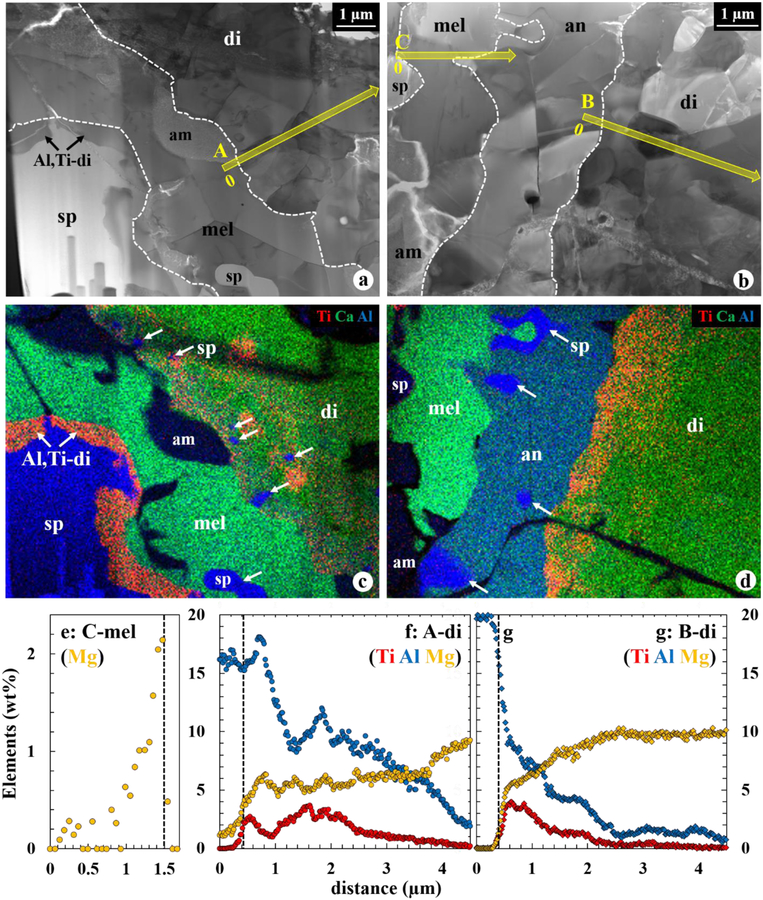
Figure 5.
BF STEM images (a, b) and corresponding combined X-ray elemental maps (c, d) in Ti (red), Ca (green), and Al (blue) of the WL rim layers from spinel to diopside, as seperated by the dashed lines. In (a, c), no anorthite is observed between melilite and diopside, and Al,Ti-rich diopside is locally present onto spinel. Fe-rich amorphous materials are present along the grain boundaries and cracks. Fine-grained spinel, indicated by arrows, are included in diopside and melilite (c) and anorthite (d). (e) Mg profile across the melilite layer, extracted from the TEM EDX spectrum images outlined in (b). The vertical dotted line in (e) represents a grain boundary between melilite and anorthite, respectively. (f, g) Al, Ti, and Mg profiles across the diopside layer, extracted from the TEM EDX spectrum images outlined in (a, b). The vertical dotted lines in (f, g) represent diopside grain boundaries with melilite and anorthite, respectively. In these profiles, 0 μm is indicated in (a, c). Diopside is zoned in Al and Ti regardless of whether or not it is in direct contact with melilite. However, diopside next to melilite contains higher Al2O3 contents and slightly lower MgO contents, compared to that next to anorthite. Abbreviation: am = Fe-rich amorphous materials.

