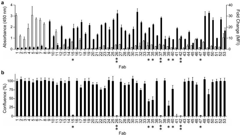
Figure 2.

Binding and function of anti-integrin-α11/β1 Fabs. (a) Binding of Fabs (x-axis) to purified integrin-α11/β1 (white bars, y-axis, left) or C2C12-α11/β1 cells (black bars, y-axis, right). Binding to purified or cell-surface integrin-α11/β1 was assessed by ELISA or flow cytometry, respectively. Data are shown for single-point measurements, and error bars indicate the standard deviation (SD) of two independent experiments. (b) Effects of Fabs (x-axis) on adhesion of C2C12-α11/β1 cells to collagen-I (y-axis). Asterisks (*) indicate Fabs that inhibited cell adhesion to collagen-I, and double asterisks (**) indicate Abs that were also characterized as full-length immunoglobulins. See Materials and Methods for details. Data are shown for single-point measurements, and error bars indicate SD of two independent experiments.
